Peripheral Artery Imaging and Intervention: A Comprehensive Guide to Angiography and Angioplasty
Peripheral angiogram and peripheral angioplasty are medical procedures used to diagnose and treat conditions affecting the peripheral arteries, which supply blood to the legs, arms, and other parts of the body outside the heart and brain.
Peripheral Angiogram
A peripheral angiogram is a diagnostic imaging test that helps visualize the blood flow in the peripheral arteries. This procedure is used to detect blockages, narrowings, or other abnormalities in these arteries. Here’s how it typically works:
- Preparation: The patient is usually asked to fast for a few hours before the procedure. A local anesthetic is applied to the area where the catheter will be inserted, usually in the groin, arm, or wrist.
- Catheter Insertion: A thin, flexible tube called a catheter is inserted into a large artery and guided to the area of interest.
- Contrast Injection: A contrast dye, which is visible on X-rays, is injected through the catheter. This dye highlights the blood vessels on the X-ray images.
- Imaging: X-ray images are taken as the dye moves through the blood vessels, allowing the doctor to see any blockages or abnormalities.
- Completion: After the images are taken, the catheter is removed, and pressure is applied to the insertion site to prevent bleeding. The patient may need to lie flat for a few hours to reduce the risk of bleeding.
Peripheral Angioplasty
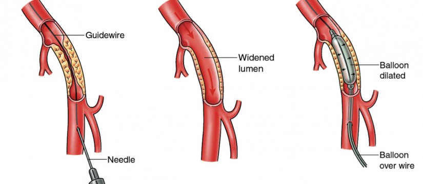
Peripheral angioplasty is a therapeutic procedure used to treat blockages or narrowings in the peripheral arteries. It often follows a peripheral angiogram if a blockage is detected. Here’s an overview of the procedure:
- Preparation: Similar to an angiogram, the patient is prepared, and a local anesthetic is administered.
- Catheter Insertion: A catheter with a deflated balloon at its tip is inserted into the artery and guided to the site of the blockage.
- Balloon Inflation: Once in position, the balloon is inflated. This presses the plaque against the artery walls, widening the artery and restoring blood flow.
- Stent Placement: In some cases, a stent (a small wire mesh tube) is placed at the site of the blockage to keep the artery open. The stent is expanded by the balloon and remains in place when the balloon is deflated and removed.
- Completion: The catheter is removed, and pressure is applied to the insertion site. The patient is monitored for a few hours or overnight to ensure there are no complications.
Risks and Benefits
Benefits:
- Minimally Invasive: Both procedures are less invasive than open surgery.
- Quick Recovery: Patients often recover faster and with less discomfort.
- Effective: Can significantly improve blood flow and alleviate symptoms like pain and cramping.
Risks:
- Bleeding: At the catheter insertion site.
- Infection: Rare, but possible.
- Allergic Reaction: To the contrast dye.
- Re-narrowing: The treated artery may narrow again over time.
- Kidney Damage: From the contrast dye, especially in patients with preexisting kidney issues.
Aftercare
After these procedures, patients are typically advised to:
- Stay Hydrated: To help flush the contrast dye from the body.
- Avoid Strenuous Activities: For a few days to allow the insertion site to heal.
- Monitor for Complications: Such as increased pain, swelling, or signs of infection at the insertion site.
Regular follow-up appointments are essential to monitor the success of the procedure and to check for any recurrence of artery narrowing.