Minimally Invasive Device Closure of Atrial Septal Defect (ASD), Ventricular Septal Defect (VSD), and Patent Ductus Arteriosus (PDA)
The closure of congenital heart defects such as Atrial Septal Defect (ASD), Ventricular Septal Defect (VSD), and Patent Ductus Arteriosus (PDA) can be accomplished using device closure procedures. These minimally invasive techniques involve the use of catheter-based devices to close the defects, reducing the need for open-heart surgery. Here’s an overview of each procedure:
Atrial Septal Defect (ASD) Closure
ASD is a hole in the wall (septum) that separates the heart’s upper chambers (atria). Device closure for ASD involves:
- Assessment: Using echocardiography and sometimes cardiac catheterization to determine the size and location of the defect.
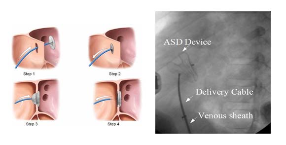
- Procedure: A catheter is inserted through a vein, usually in the groin, and guided to the heart. The closure device, which resembles a small umbrella or disc, is then deployed to cover the defect.
- Device: The most commonly used devices are the Amplatzer Septal Occluder and the Gore Helex Septal Occluder.
Ventricular Septal Defect (VSD) Closure
VSD is a hole in the wall separating the lower chambers (ventricles) of the heart. Device closure for VSD includes:
- Assessment: Echocardiography and cardiac catheterization are used to visualize the defect.
- Procedure: Similar to ASD closure, a catheter is inserted through a blood vessel and guided to the heart. The closure device is positioned to cover the VSD.
- Device: Devices like the Amplatzer VSD Occluder are used.
Patent Ductus Arteriosus (PDA) Closure
PDA is a persistent opening between the aorta and the pulmonary artery that normally closes after birth. Device closure for PDA involves:
- Assessment: Diagnosis through echocardiography to measure the PDA.
- Procedure: A catheter is introduced through a vein or artery and advanced to the site of the PDA. The closure device, often a coil or a plug, is deployed to block the abnormal blood flow.
- Device: The Amplatzer Duct Occluder and coils are commonly used for PDA closure.
Advantages of Device Closure
- Minimally Invasive: These procedures are less invasive than traditional surgery, requiring only small incisions or needle punctures.
- Shorter Recovery Time: Patients typically recover faster and with less discomfort compared to open-heart surgery.
- Reduced Risk: Lower risk of complications such as infection and bleeding.
Considerations and Follow-Up
- Patient Selection: Not all defects are suitable for device closure. The size, location, and anatomy of the defect, as well as the patient’s overall health, are considered.
- Follow-Up: Regular follow-up with echocardiography is necessary to ensure the device is in place and functioning correctly. Monitoring for potential complications like device migration or residual shunting is also important.
Conclusion
Device closure of ASD, VSD, and PDA represents a significant advancement in the management of congenital heart defects, offering a safer and less invasive alternative to traditional surgical methods.